The endogenous fluorescence of proteins is mainly derived from aromatic amino acids such as tryptophan, tyrosine and phenylalanine. In water, the maximum excitation light wavelength of tryptophan is around 270-280 nm and the peak wavelength of its emitted light is close to 350 nm. The emission spectrum of the protein is very sensitive to the polarity of the solvent and the external environment. When the protein is denatured, the tryptophan residue is exposed to water and its emission wavelength becomes longer. This change in peak emission wavelength can be used to detect denaturation of proteins. Here, we show how to use the SpectraMax® iD3 Multi-Purpose Plate Reader to detect endogenous tryptophan fluorescence. The tryptophan standard curve was used to show the high sensitivity of the detection method, while the shift in the tryptophan emission peak was detected by a lysozyme denaturation experiment. Lysozyme contains two tryptophan residues and therefore emits fluorescence when exposed to ultraviolet light. When lysozyme is degraded, the peak of the emitted light of tryptophan is shifted by 6-10 nm. We can perform fluorescence spectral scanning of lysozyme before and after degradation to capture and detect the peak shift that occurs. The SpectraMax® iD3 Multi-Purpose Plate Reader combines the high sensitivity and wavelength scanning capabilities required for endogenous tryptophan fluorescence detection. In addition, SoftMax® Pro software automatically identifies the best excitation and emission wavelengths through the Spectral Optimization Wizard, while automatically calculating the offset of the emission peaks for subsequent analysis.
Method We prepared a sample of tryptophan with a starting concentration of 100 μM and diluted it. The original concentration and the diluted sample were added to a UV-transparent 96-well plate in the form of three replicate wells per sample (200 microliters per well). The lowest limit of detection (LLD) was calculated by adding PBS as a control to the six blank wells. The Spectral Optimization Wizard of SoftMax Pro software is optimized to achieve the best excitation and emission wavelengths. Through the results of wavelength optimization, we can accurately detect the ratio of dilution of tryptophan. We treated 10 mg/mL lysozyme solution with a final concentration of 5 M guanidine hydrochloride to denature it. Then we used a SpectraMax iD3 plate reader to perform spectral scanning on the lysozyme sample before and after denaturing. The excitation light was 270 nm and the emission was Between 300nm and 450nm. The emitted light peaks are automatically identified using a pre-programmed program in SoftMax Pro Software.
Materials • SpectraMax iD3 Multi-Purpose Plate Reader
(Molecular Devices cat. #ID3-STD)
• L-tryptophan (Sigma cat. #T-0254)
• Lysozyme (Sigma cat. #L6876)
• Hydrazine hydrochloride (Sigma cat. #G7294)
• UV high permeability 96-well plate
(Greiner cat. #655801)
• Phosphate buffer (PBS), pH 7.4)
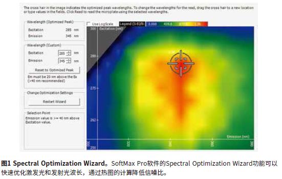
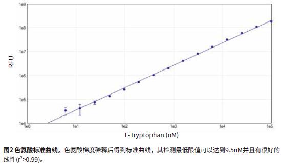
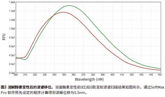
The Spectral Optimization Wizard for SoftMax Pro software optimizes the excitation and emission wavelengths (Figure 1). This feature allows the measurement of these wavelengths to have an optimum signal-to-noise ratio (excitation wavelength 285 nm, emission wavelength 345 nm). By optimizing the wavelength, the SpectraMax iD3 reader can accurately measure the standard curve of tryptophan with a minimum detection limit of 9.5 nM and good linearity (r2 > 0.99) (Figure 2). The spectral scanning results showed that the peak wavelength of the emitted light after lysozyme degradation migrated from 350 nm to 355.5 nm with a slight increase in fluorescence intensity (Fig. 3). The extent of the peak wavelength of this emitted light is also very close to the expected value.
in conclusion
The SpectraMax iD3 reader can characterize proteins by measuring the endogenous fluorescence of tryptophan. Using a bandwidth of 15 nm excitation light and 25 nm emission light, the reader can detect low concentrations of tryptophan in the low wavelength spectral range. In addition, the Spectral Optimization Wizard automatically recognizes the optimum excitation and emission wavelengths. Using the pre-programmed procedures of SoftMax Pro software, researchers can automatically detect peak wavelengths and their migration before and after lysozyme degradation, saving a lot of time for data analysis.
nucleic acid extractor specifically adsorbs nucleic acid molecules in samples through magnetic beads, and then adsorbs transfers magnetic beads through the movement of magnetic rods in different holes, so as to achieve the purpose of purifying nucleic acid molecules. Former 1B48 nucleic acid extractor instrument can automatically extract nucleic acid from whole blood, bacteria, plasmid, virus, serum free and plant samples, widely used in scientific research, CDC, food safety, clinic testing, forensic medicine, and other fields.
Nucleic Acid Extrator,Dna Nucleic Acid Extrator,Nucleic Acid Extrator For Lab,Automated Nucleic Acid Extrator
Nanjing Superyears Gene Technology Co., Ltd. , https://www.superyearsglobal.com