In recent years, with the development of single-molecule fluorescence imaging technology of living cell system, the study of membrane protein single molecule, especially receptor kinetics, has become one of the most active research directions in the field of single molecule research. The super-resolution imaging technology developed in recent years has higher resolution and higher positioning accuracy than traditional optical microscopes because it can break through the optical diffraction limit.
The Nanoimager, the latest super-resolution fluorescence microscope from Oxford Nanoimaging, developed by the team of professors of the University of Oxford, Achillefs Kapanidis, is the world's first large-field single-molecule FRET microscope with super-resolution at a single molecule. Research fields such as tracing, live cell imaging, protein interaction, and 3D imaging play an important role.
♦ Horizontal resolution <20nm; vertical resolution <50nm
♦ Stability: <1 μm/K drift; <1 nm (1 Hz to 500 Hz) amplitude ♦ Supports simultaneous two-color imaging and sequential four-color imaging ♦ Class 1 laser, safe to use
1, single molecule FRET
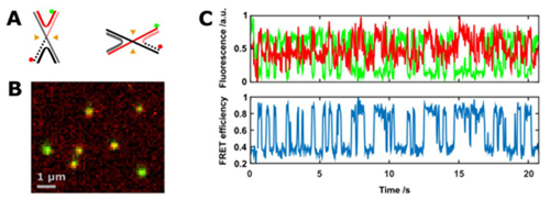
The Nanoimager directly reflects the diffusivity and estimated size of the fluorescent particles in the purified sample, with sensitivity (single fluorescent molecular level) and specificity (two-color labeling can significantly reduce the possibility of detecting impurities).
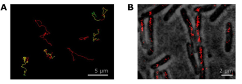
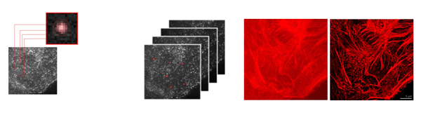
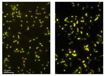
Each imaging channel of the Nanoimager has a large field of view of 50 μm x 80 μm and is evenly illuminated for high-throughput imaging of single molecules or cells and rapid data collection. Figure 4 shows imaging of different phenotypes of mutant E. coli cells at a rate 10 times faster than other techniques. In order to obtain reliable results for different phenotypes, a large number of cells need to be compared. Using a Nanoimager with a large field of view that autofocuses and automatically acquires data can dramatically speed up the entire experiment with speed and throughput. Combining large field of view with super-resolution imaging is a unique advantage of the Nanoimager.
1. Material of our mildew tape
1) Transparent acrylic adhesive + polyester, thickness: 0.5mm and 0.8mm
2) PVC + acrylic adhesive, thickness is about 1.5mm, different colors for choosing.
3) PE + butyl adhesive, long serfice life and and good temperature resistance.
2. Features of our mildew tape
1) Good adhesion and soft material, can be adhered to many different surfaces.
2) Waterproof, resist the water, oil and dust go inside the cracks and corners, resist mildew.
3) Long service life, easy to clean.
4) Different colors and patterns for choosing.
5) Easy to operate, Do not need to use other complicated tools, the waterproof tape is affixed around the sink, use PE Self Adhesive, repeatedly press several times.It adhesive sticks well can seal the gap between the tile and the wall.
3. Applications of our mildew tape
Widely used for sealing the seams of gas stove, sink, basin, bathtub and walls, it could prevent them from getting mouldy and black, keep your kitchen and bathroom clean and new for a long time.
Mildew tape,Anti-mildew tape,anti mildew tape,caulking tape,caulking strip,caulk strip,caulk tape
Kunshan Jieyudeng Intelligent Technology Co., Ltd. , https://www.jerrytapes.com