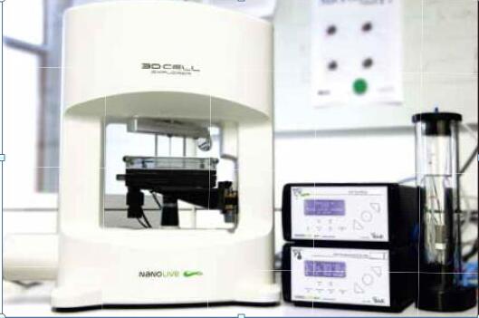
New Technology for Nematode Research ----NanoLive Real-Time Label-Free 3D Microscope
The importance of 3D cell Explorer real-time label-free technology for C. elegans live cell imaging
C. elegans is one of the most widely used model organisms in biomedical research. A major problem with current imaging techniques is phototoxicity, which leads to interference with live cell dynamics observations, and the development of new label-free, high-resolution imaging is critical to better understanding the anatomy and physiology of this precious organism. important.
Here, we show a video of unmarked imaging of C. elegans with 3D cell explorer, how to overcome the above problems. First, the 3D cell explorer illuminates the sample 100 times smaller (~0.2nW/m2) than the light microscopy (~1nW/m2) as a reference, which greatly reduces the damage to living cells with a resolution of up to 200 nm. Therefore, high-resolution and high-frequency imaging of nematode live cells can be performed.
The videos and images below are the results of Friedrich Puruser and Nikita Vladimirov from the Max de Bruk Centre for Molecular Medicine in Berlin.
Https://v.qq.com/x/page/f1351nryoj5.html
3D cell Explorer can image C. elegans at different stages of embryonic development
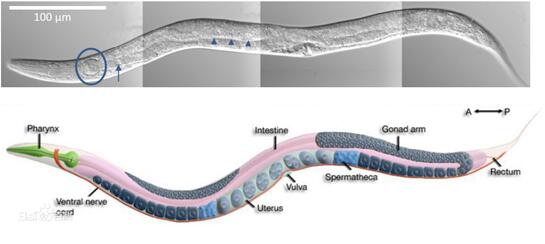
The video shows the 3D cell Explorer's real-time marker tomographic 3D scanning process for different stages of early development of Caenorhabditis elegans: A. Unfertilized egg cells in the uterus (green arrow on the right) and sperm (green arrow on the left); C. Embryonic stage AB: The membrane between the two cells is marked with a green arrow; D. The embryonic phase AB 2 : The embryo consists of 4 cells. Egg shells are clearly visible, indicated by green arrows; E. Embryonic phase AB n : Embryos consist of many (n) cells. The maternal mouth can also be seen in the field of view, as indicated by the green arrow; F. There are two different embryos in the uterus that develop at different speeds: AB 2 at the bottom and AB n at the top.
Https://v.qq.com/x/page/r135145lmr4.html

3D cell Explorer for unmarked high definition imaging of the C. elegans reproductive system
The figure below shows that unfertilized oocytes stay in the ovaries and are transferred one by one through the seminal vesicles to the uterus where they meet and fertilize the sperm.

3D Cell Explorer's outstanding advantages in online pest research
* Holographic imaging of a 360-degree rotating light source;
* Non-invasive or without staining marker imaging;
* No damage: low power laser, RI-based, damage-free real-time 3D cell structure imaging;
* Arbitrary digital coloring : up to 7 colors;
* Nano-scale resolution : XY=200nm;
* Ultra-high imaging speed: 2 seconds of fast 3D holographic imaging;
*High throughput: integrated 3-channel fluorescence, up to 10 color markers (7DI+3 color fluorescence)
Seasoned Scallop,Seasoned Sea Scallop,Seasoned Spicy Scallop,Seasoned Scallop Lips
DALIAN HAIBAO FOODS CO., LTD. , https://www.haibaoseafoods.com