Introduction
Advantage · Rapid imaging to efficiently identify monoclonal cell lines · Accurately track the growth of individual cells · Can image cells in semi-solid medium or liquid medium, flexible selection of experimental protocols |
The regulatory standards for biopharmaceutical approvals have been constantly changing. Regulatory regulations for the monoclonal source of cell lines have driven us to use more efficient techniques or methods in biopharmaceutical development. Many researchers typically use imaging systems (such as CloneSelect Imager) to verify the growth of monoclonal and monitored cells in liquid media. In fact, the CloneSelect Imager system can also image cells in semi-solid media. The location of the cells in the semi-solid medium is relatively fixed, allowing researchers to more accurately image the same cell at different time points.
Conventional methods in the laboratory typically use limited dilution for cell cloning. However, this method uses liquid media to culture cells, making tracking cells difficult or less accurate. Because of the ease of movement of the suspended cells within the well during processing of the microplate (Figure 1). It is not possible to ensure that the same cell is imaged at different time points, thus greatly reducing the accuracy of monoclonal validation. In addition, limited dilution has the potential to add multiple cells in one well, requiring additional multiple subclones to increase the probability of monoclonal.
The use of semi-solid medium to culture cells provides researchers with a faster and simpler method of cloning and is useful for tracking cell growth. The position of the cells in the semi-solid medium is relatively fixed, the cell position does not move during routine experimental procedures, and can be grown into individual cell clones. Therefore, multiple cells can be planted in the same well, and imaging at different time points can clearly track the growth of individual cells and provide monoclonal digital verification data.
CloneSelect Imager provides researchers with a way to image cells in semi-solid media. This system uses a non-invasive assay that allows for accurate quantification of cell growth without the need for labeled cells for direct imaging; single-cell source cloning validation ensures that only stable monoclonals are selected for downstream studies. In this paper, the imaging performance of CloneSelect Imager in suspension CHO-s cells in CloneMediaTM CHO Growth A semi-solid medium was verified by practical experiments. This semi-solid medium component is specifically optimized for growth in a variety of CHO cell lines.
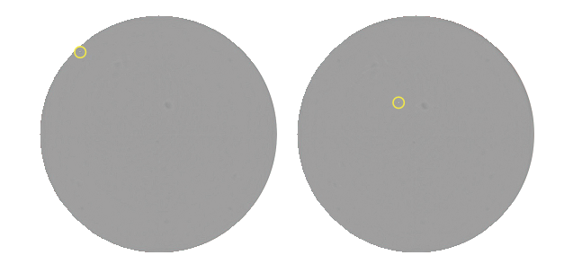
Figure 1: Limited-dilution plated 96-well plate with same well CHO-s cell imaging image: The microplate was imaged every 8 hours using the CloneSelect Imager system. Just a routine experimental procedure, the cells have moved between the two images, and the yellow marks the location of the cells.
Experimental operation flow
The flow of CHO-s cell culture and imaging is shown in Figure 2.
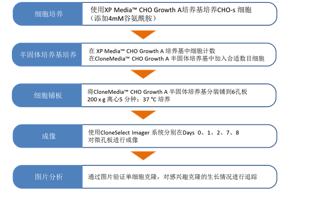
Figure 2: General flow chart for tracking individual cell growth experiments using CloneMedia CHO Growth A semi-solid medium. XP Media CHO Growth A and CloneMedia CHO Growth A media provide a stable CHO cell culture, cloning and screening method; imaging with CloneSelect Imager allows for monoclonal validation and cell growth tracking.
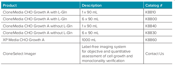
CHO-s cells were first cultured using XP MediaTM CHO Growth A liquid medium (Cat. No. K8860), and 4 mM glutamine was added before the culture, and cultured at 37 °C. CHO-s cells were then plated into semi-solid medium CloneMedia CHO Growth A (Cat. No. K8810) with a plating density of 500 cells/ml. The cells were mixed and dispensed into a 6-well plate (Greiner catalog # 657185) at 2 ml/well. At the same time, 10 uM diameter sterile beads (Thermo Scientific catalog # G1000B) can also be added to the semi-solid medium as a reference for the imaging position. The addition of microbeads is not an essential step in the above CHO-s semi-solid culture process.
Among them, the A1 well should be added with 2ml 50000cells/ml cell suspension as the reference hole for focusing, which is convenient to determine the correct focus value. 2 ml of 500 cells/ml cell suspension was added to each of the other wells, and then centrifuged at 200 xg for 5 minutes and cultured at 37 °C.
CHO-s cell imaging
Microplates were imaged at multiple time points using CloneSelect Imager to provide digital evidence of monoclonal images and to track cell growth. Each plate was imaged at several time points of Day 0, 1, 2, 7, and 8. For Day 0 imaging, the microplates were first incubated at 37 °C for 4 hours. When imaging, first use the autofocus function for the A1 hole, the software automatically finds the optimal focus value, and obtains the optimal imaging effect of the whole board (Figure 3). Moreover, this focus value is automatically recorded and the same focus value is used for the imaging process at other points in time.
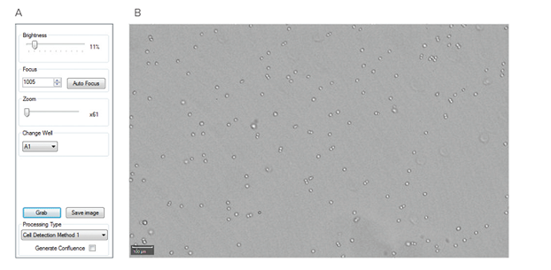
Figure 3: CloneSelect Imager system sets the focus value (A) focus setting software interface, Auto Focus button is used to automatically set the optimal focus value for 6-hole CHO-s cell imaging. (B) A1 hole cell imaging image obtained using the Auto Focus function.
result
CHO-s cells can be grown in CloneMedia CHO Growth A semi-solid medium to form compact spherical clones that are very easy to identify. The cell density was 500 cells/ml, and the number of individual clones formed per well was clearly identifiable on Day 8 (Figures 4A and 4B). The use of semi-solid media facilitates the growth of multiple independent clones in a single well, with relatively limited dilution, requiring a significant savings in the number of microplates and media.
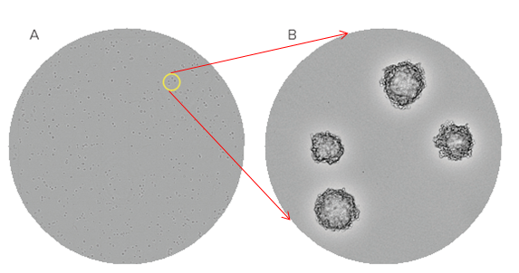
Figure 4: CHO-s cells in CloneMedia CHO Growth A semi-solid medium obtained by CloneSelect Imager system image: (A) Whole-hole images obtained from Day 8 days of CHO-s cells with a planting density of 500 cells/ml. (B) A magnified picture of a partial clone in the well, which clearly distinguishes a single clone. The yellow circle (A) indicates the portion of the enlarged view of Figure B.
CloneSelect Imager software can be used to zoom in on individual clones, and by linking cloned images at different time points, the growth of the clones can be tracked to verify whether the clones are monoclonal. As shown in Figure 5, a picture of two clones is displayed. By comparing the growth of the two clones at different time points and tracking to the Day 0 day image, it can be determined whether the clone grows in single or multiple cells. Additional microbeads (yellow circle markers) added to the semi-solid medium can be used as a reference for imaging locations to determine that the same clone is imaged each time.

Figure 5: CloneMedia CHO Growth A CHO-s cells in semi-solid medium. Picture: CloneSelect Imager images 6-well plates at multiple time points. In Day 0, as shown in the figure, it can be clearly observed that the above picture is a cell, and the figure below shows two cells. The yellow circle marks the position of the beads and is used as a position reference to determine that the same clone is used for each imaging.
in conclusion
The use of CloneMedia CHO Growth A semi-solid medium to culture CHO cells is a more efficient and accurate cloning method than limiting dilution with liquid medium. The semi-solid medium can fix the position of the cells and does not affect the positional movement of the cells during routine experimental procedures. This method can more accurately track the growth of a single cell into a clone. Although CloneSelect Imager is commonly used for imaging cells in liquid media, it can also be used to image cells in semi-solid media, providing a more accurate tool for tracking cell growth and verifying monoclonal.
Supplier Extract Powder
We're Professional Supplier Extract Powder manufacturers and suppliers in China specialized in providing high-quality products at low price. We warmly welcome you to buy or wholesale bulk Supplier Extract Powder for sale here from our factory. For a free sample, contact us now.
Supplier Extract Powder,Supplier Extract ,Supplier Powder Manufacturer in China
Shaanxi Kang New Pharmaceutical co., Ltd. , https://www.apipepdites.com